
We've come a long way in visually diagnosing endometriosis, and in 2026, a new potential imaging agent is on the horizon. But first, let's briefly review the historical development of detecting endometriosis.
From Autopsy to Microscopy
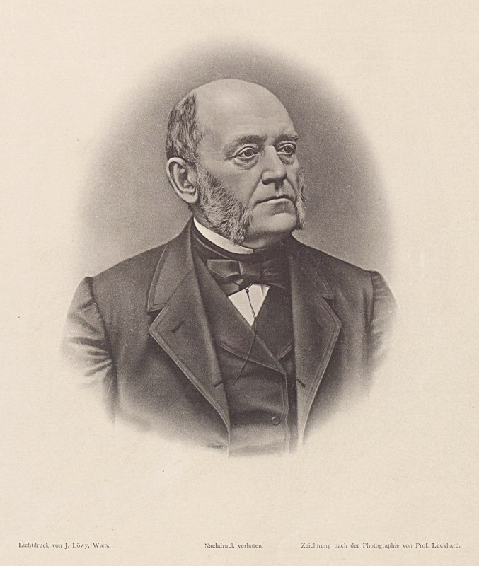
The primary, definitive visual, and microscopic diagnosis of endometriosis is attributed to the Austrian pathologist Carl Freiherr von Rokitansky in 1860. He is known for single-handedly transforming pathology from speculative philosophy into an exact, clinical science.
Over his career, he personally performed or supervised over 30,000 to 60,000 autopsies, giving him unprecedented insight into human anatomy. Rokitansky did not set out to find endometriosis; rather, his discovery was a byproduct of his relentless, systematic autopsy routine at the General Hospital of Vienna.
He regularly encountered female cadavers with severe pelvic adhesions, cysts, and unexplained growths in the abdomen. Using a microscope on these autopsy tissue samples, Rokitansky noticed something extraordinary: abnormal growths on the ovaries, bladder, and intestines that contained cellular structures identical to the glands and stroma of the endometrium (the lining of the uterus). He documented the tissue and categorized it as tumor-like, because the growths appeared aggressive and invasive to him.
In fact, the current definition of endometriosis still reflects this paradox: benign tissue acting destructively outside its "usual home". Today, the neuro-immunological correlations behind this behavior are becoming increasingly clear.
Early diagnosis from the 19th century on relied on naked-eye observation (macroscopic) of surgical specimens, often called "adenomyoma" or "chocolate cysts".
Ultrasound Enters the Picture
Ultrasound officially entered gynecology and obstetrics in 1958. The original technique relied on a giant, primitive transabdominal machine adapted from industrial metal-testing and naval sonar equipment. It successfully differentiated between solid tumors and benign fluid-filled ovarian cysts.
Ultrasound brought a new layer of visual detection, although it still often relies heavily on the user's pattern recognition and experience (radiologist or OB-GYN) and on the machine's resolution or software capabilities.
Mapping Deep Disease
The current gold standard for 3D mapping and pre-surgical planning of complex pelvic endometriosis is Advanced Transvaginal Ultrasound (TVS) incorporating the IDEA (International Deep Endometriosis Analysis) protocol, coupled with 3D Volume Render/VOCAL processing. All potential lesions are noted, sparing time during anesthesia if a laparoscopic excision is performed.
While traditional 2D ultrasound provides real-time movement data, specialized 3D transvaginal volume transducers upgrade the mapping quality via:
- Coronal Plane Reconstruction: Standard 2D ultrasound cannot easily see the coronal view. 3D ultrasound mathematically reconstructs this plane, allowing precise structural analysis of the uterosacral ligaments and the rectovaginal septum.
- VOCAL (Virtual Organ Computer-Aided Analysis): This specific software maps out and tracks the precise volumes of complex, irregular deep endometriotic nodules over time.
- Quantitative Gray Value Analysis: 3D tissue software helps evaluate tissue density to differentiate between purely fibrous, rigid tissue and vascularized active lesions.
MRI and the Full Pelvic Map
In the early 1990s, pelvic magnetic resonance imaging (MRI) transformed endometriosis management by providing a comprehensive, non-invasive map of the entire pelvis. It bridged the gap between initial screening and the operating room. In 1999 pioneering work by researchers like K. Kinkel established specific MRI characteristics for deep infiltrating endometriosis (DIE), shifting MRI from an ovarian cyst tool to a comprehensive deep tissue mapping modality. Since endometriosis bleeds internally with each menstrual cycle, these locked-in blood products act as natural "contrast agents" that light up under specific sequences in the MRI.
Today, MRI is classified as the ultimate second-line problem-solving tool. If a transvaginal ultrasound is inconclusive, or if a patient has a highly symptomatic profile but normal ultrasound results, a specialized pelvic MRI is ordered.
In summary: While 3D TVS maps deep infiltrative disease and ovarian cysts with accuracy comparable to or exceeding MRI, it still struggles to reliably visualize superficial peritoneal endometriosis (flat surface implants). And this is where the new imaging agent comes in.
New imaging agent Maraciclatide
Maraciclatide (formerly NC100692) is a synthetic radiolabeled tracer that specifically binds to αvβ3 integrin receptors, which are overexpressed in angiogenesis and inflammation. The compound, developed by Serac Healthcare, is being investigated using SPECT/CT for the non-invasive imaging of conditions such as endometriosis, interstitial lung diseases (ILD), and various types of cancer. It is administered intravenously and visualized using gamma scintigraphy or SPECT-CT.
Good Results, Hope & Potential for Lesion Detection
In April 2026, Serac Healthcare and the University of Oxford published Phase II data showing that 99mTc-maraciclatide, a molecular imaging agent, can visualize endometriosis non-invasively. The DETECT study showed the agent's great potential for the non-invasive visualization of superficial peritoneal endometriosis, which could reduce the need for diagnostic surgery in this most prevalent subtype of endo. This is important, since peritoneal endometriosis has historically been hard to detect via ultrasound or MRI. It accounts for 80% of all laparoscopically diagnosed endometriosis, making a non-invasive detection method particularly significant.
Within the study, endometriosis was detected in 14 of 17 surgically positive participants. Notably, 10 participants with superficial peritoneal endo (SPE) had undergone conventional imaging (TVS and/or MRI) within the previous 12 months, and none of those scans had detected their SPE. No false positives were reported in this small study, corresponding to 100% specificity. Lesions were detected across all endometriosis subtypes, including two cases of thoracic endometriosis, regardless of current treatments.
Beyond diagnosis, this agent gives hope for monitoring disease recurrence and measuring treatment response as well. If these Phase II results are reproduced in the Phase III studies, maraciclatide has the potential to be an extremely valuable tool for researchers and patients alike.
Side Effect Profile
Maraciclatide differs fundamentally from gadolinium-based contrast agents (GBCAs), which are used in MRI enhancement, in both its chemical composition and its safety profile. To date, there is no evidence that maraciclatide causes serious long-term effects such as nephrogenic systemic fibrosis (NSF) or tissue deposits, which are typically associated with gadolinium.
Maraciclatide is a small synthetic peptide tagged with technetium-99m, a radioactive tracer used in nuclear medicine for decades. Technetium-99m has a physical half-life of about six hours, and in a Phase I study the maraciclatide component had a terminal elimination half-life of about one hour.
Unlike gadolinium-based MRI contrast agents (which do not emit radiation during MRI), maraciclatide is a radiopharmaceutical used in SPECT/CT. The main risk here is low radiation exposure, which is comparable to that of other common nuclear medicine examinations and is considered acceptable for diagnostic use. How long it stays in the body depends on where it distributes after injection.
In Phase I studies, the agent was safe, with mild, self-limiting side effects. The dosimetry is similar to that of other 99mTc agents. However, long-term data are not yet available.
Outlook
The findings of this study could reduce the number of women undergoing unnecessary laparoscopic or thoracoscopic surgery while improving the accuracy and speed of diagnosis, as well as disease monitoring and management for millions of women across the world.
Further validation is required before this technique can be used in routine clinical practice. International multi-centre Phase III studies are due to begin later this year.